Aug 01, 2025
3D Ultrasound: How It Enhances Accuracy in Modern Diagnostics
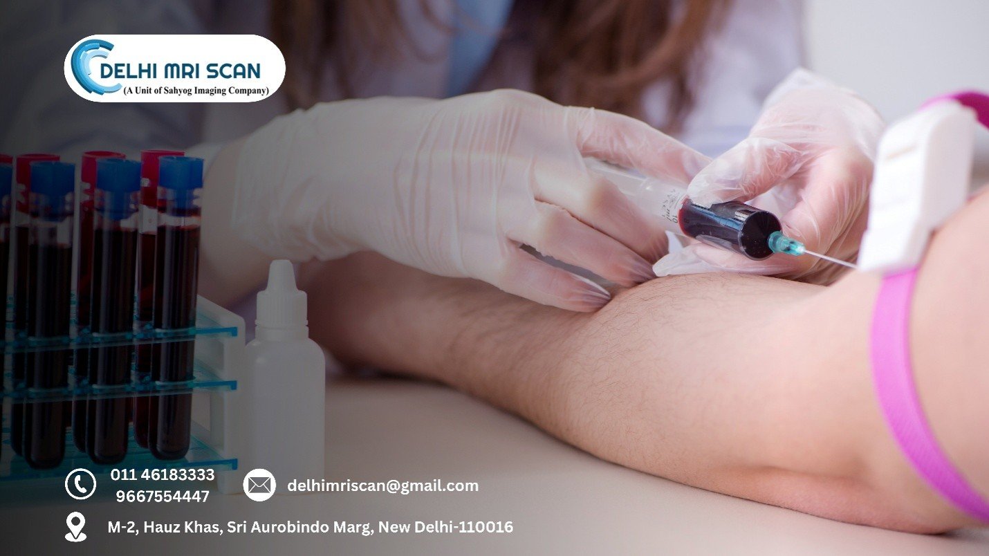
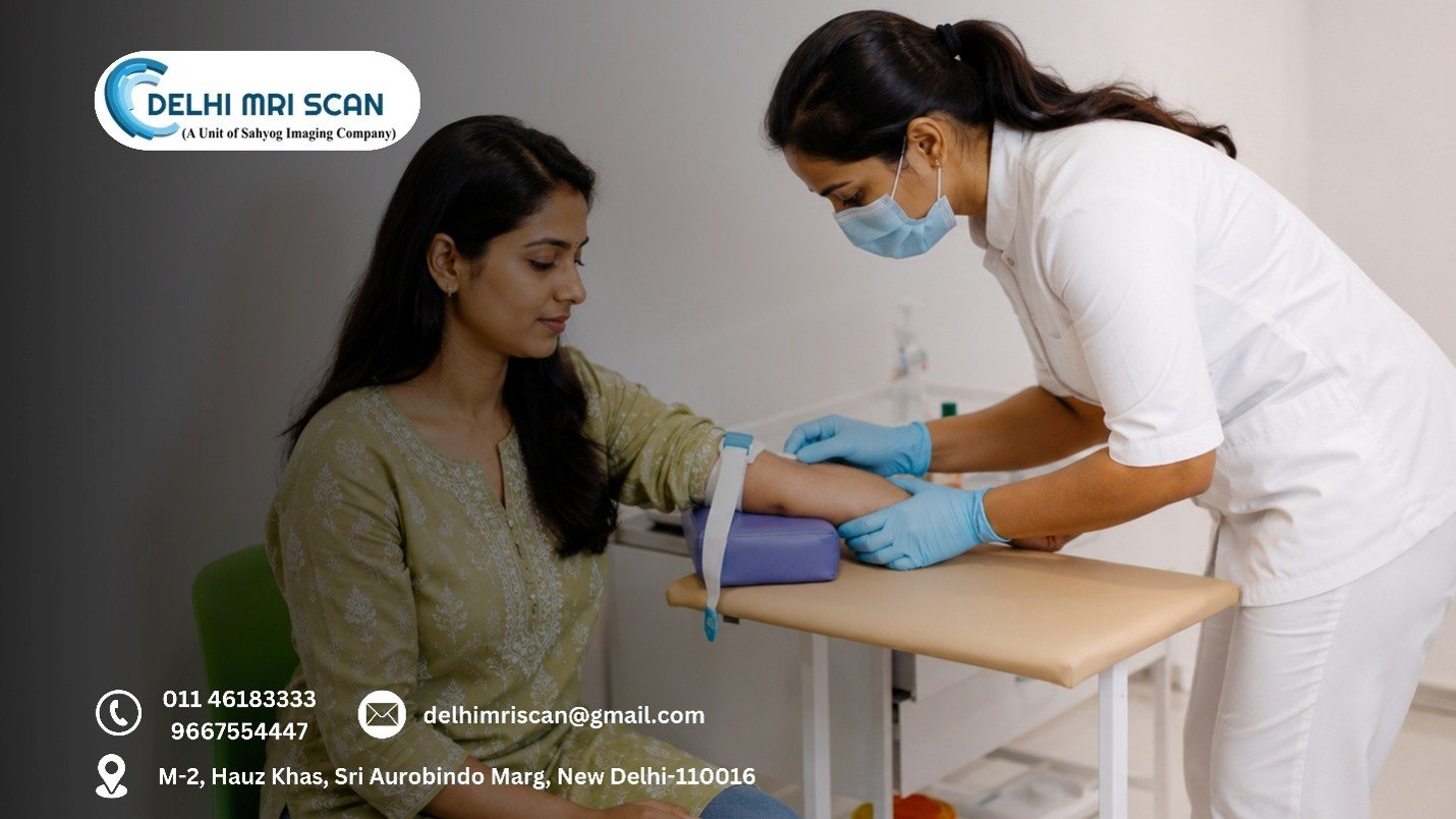
Patients and physicians alike expect accuracy, speed, and dependability in diagnostic techniques in the rapidly changing healthcare environment of today. With the help of advanced imaging technologies, Delhi MRI Scan can more clearly interpret medical conditions in addition to providing an inside look at the body. Among these innovations,
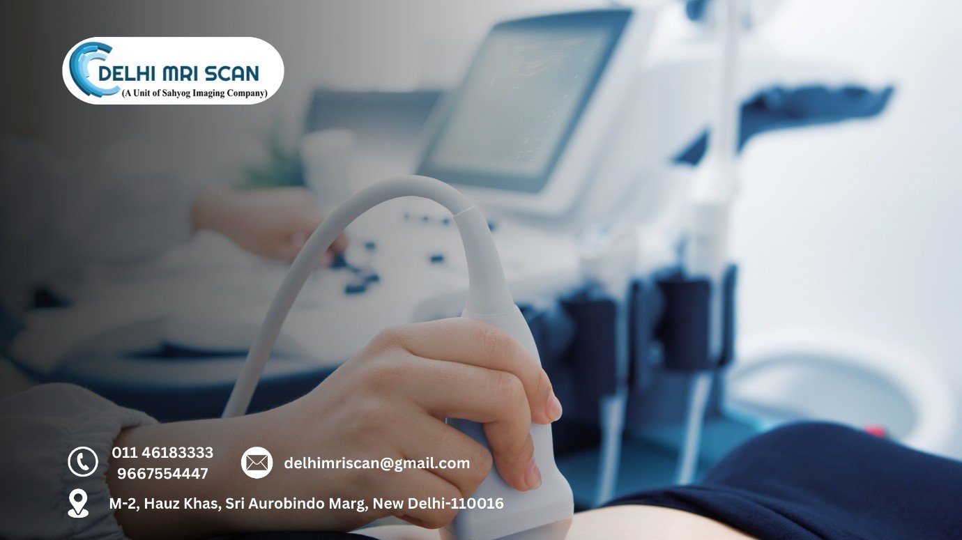
3D ultrasound stands out as an exceptional instrument that has revolutionized the practice of contemporary diagnostics. 3D ultrasound is revolutionizing medical imaging because it provides depth, detailed visualization, and more accurate information than traditional two-dimensional scans.
The Evolution of Ultrasound Technology
One of the most popular and safest diagnostic methods for a long time has been ultrasound. At first, 2D ultrasound was the norm, giving medical professionals flat, cross-sectional pictures of tissues, internal organs, or a growing foetus. Despite their effectiveness, these pictures occasionally left space for ambiguity and frequently required interpretation skills.
Volumetric imaging became available to the medical community with the advent of 3D ultrasound, allowing doctors to view three-dimensional structures in real time. This development not only made visualization better but also significantly increased the accuracy of 3d ultrasound, aiding medical professionals in more efficient diagnosis, monitoring, and treatment planning.
What Makes 3D Ultrasound Different?
Capturing data is the primary distinction between 2D and 3D ultrasound. 3D technology records a large amount of data that can be assembled into a full three-dimensional image, whereas a 2D ultrasound creates flat images of slices of anatomy.
This implies:
- Organs, blood vessels, and foetal development are among the structures that can be observed in greater detail.
- Physicians can rotate and manipulate images to view from multiple angles.
- There’s less dependence on guesswork or interpretation, improving the accuracy of 3d ultrasound significantly.
Applications of 3D Ultrasound in Modern Diagnostics
1. Obstetrics and Gynaecology
Prenatal care is arguably the application of 3D ultrasound that is most well-known. Beyond the emotional joy that expectant parents get from viewing lifelike pictures of their unborn child, the technology has a vital medical function.
Physicians are better equipped to monitor placental function, evaluate the amniotic environment, detect congenital abnormalities, and evaluate foetal growth. The accuracy of 3d ultrasound gives parents and medical professionals peace of mind by guaranteeing early intervention in the event of any developmental concerns.
2. Cardiology
Cardiovascular imaging now relies heavily on 3D ultrasound. Subtle variations in heart structure and blood flow can occasionally go unnoticed by conventional 2D scans. Cardiologists can examine heart chambers, valves, and vascular structures in unprecedented detail with 3D.
This is especially helpful for planning interventional procedures, tracking post-operative recovery, and diagnosing congenital cardiac defects. The accuracy of 3d ultrasound enhances patient outcomes and lowers misdiagnosis by offering practical insights.
3. Oncology
Accuracy is crucial for both cancer diagnosis and treatment planning. Oncologists can measure tumours precisely, see them more clearly, and track their growth or reaction to treatment with 3D ultrasound.
This improved imaging reduces needless procedures and supports targeted therapies. By improving the accuracy of 3d ultrasound, more effective treatment customization by oncologists results in improved patient quality of life and survival rates.
4. Musculoskeletal Imaging
In order to properly diagnose musculoskeletal issues, which range from sports injuries to degenerative joint conditions, detailed imaging is frequently necessary. High-resolution pictures of muscles, tendons, and joints are made possible by 3D ultrasound, which facilitates the detection of abnormalities, inflammation, and tears.
Ultrasound provides superior diagnostic detail but is quicker, easier to obtain, and less expensive than MRI. The improved accuracy of 3d ultrasound enables orthopaedic specialists to create accurate surgical and rehabilitation plans.
5. Interventional Procedures
The way minimally invasive procedures are carried out has changed as a result of 3D ultrasound. The ability to see in three dimensions makes interventions safer and more successful, whether it's performing targeted therapy, guiding a biopsy, or inserting a catheter.
Real-time guidance with fewer complications is advantageous for radiologists and surgeons because of the improved accuracy of 3d ultrasound.
Advantages of 3D Ultrasound Over Traditional Imaging
Beyond applications, the switch to 3D ultrasound has several advantages that make it essential for diagnosis:
- Non-invasive and safe: It avoids radiation exposure by using sound waves, just like conventional ultrasound.
- Real-time imaging: Offers a dynamic evaluation of tissues and organs.
- Patient-friendly: Easy, quick, and very educational.
- Improved collaboration: Clear 3D images can be shared between doctors and patients, enhancing trust and communication.
- Cost-effective: Less expensive than CT or MRI scans, but in some circumstances, they have similar diagnostic value.
The Future of 3D Ultrasound
3D ultrasound appears to have an even brighter future thanks to developments in machine learning and artificial intelligence. Future developments already include improved resolution, automated image interpretation, and integration with additional diagnostic tools. As technology develops further, the accuracy of 3d ultrasound will only improve, making it an even more vital part of modern healthcare.
Final Summary
3D ultrasound is a revolution in diagnostic imaging, not just a technical advancement. It fills the gap between detection and accurate diagnosis by providing realistic, accurate, and multi-dimensional views of the human body. Its uses are numerous and significant, ranging from cardiology and oncology to prenatal care and beyond. For patients, this translates into more dependable care, safer processes, and quicker responses. It signifies confidence in clinical decision-making for physicians. Additionally, it represents a step toward patient-centred, precision medicine for the healthcare sector. These developments at Delhi MRI Scan in Hauz Khas demonstrate the dedication to using state-of-the-art technology to improve results and make sophisticated diagnostics accurate and available to all.
Related post
Dec 15, 2025
Tests Every Woman Should Consider After 30
Dec 02, 2025
How to Choose the Right Diagnostic Centre

Nov 17, 2025