Oct 15, 2024
The Importance of Echocardiography: A Critical Tool for Heart Health Evaluation
When it comes to diagnosing heart conditions, one of the most effective and non-invasive tools is echocardiography, commonly known as an "echo." This imaging technique uses sound waves to create detailed images of the heart's structure and function, helping healthcare professionals evaluate the heart's size, shape, and pumping ability. In this blog, we will explore what echocardiography is, its types, how it works, why it’s important, and what to expect during the procedure.
What is Echocardiography?
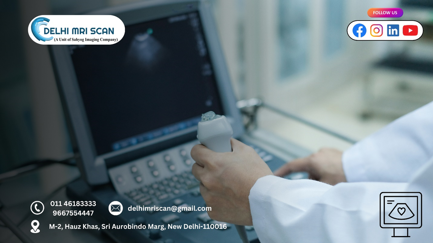
Echocardiography is a diagnostic test that uses high-frequency sound waves (ultrasound) to produce images of the heart. It provides real-time images of the heart's chambers, valves, and blood flow, enabling doctors to assess various aspects of cardiac health. This non-invasive procedure is often used to diagnose and monitor heart diseases and conditions, such as heart valve problems, heart failure, congenital heart defects, and cardiomyopathy. For those seeking the best echocardiography test in Hauz Khas, this imaging method is a top choice.
Types of Echocardiography
Echocardiography comes in several different forms, each tailored to specific diagnostic needs:
- Transthoracic Echocardiography (TTE): This is the most common type of echocardiogram. During a TTE, a technician places a transducer (a small device that emits sound waves) on the chest to capture images of the heart. It’s a simple, painless procedure that usually takes 30 to 60 minutes.
- Transesophageal Echocardiography (TEE): In this procedure, a thin tube with a transducer at the end is inserted through the mouth and into the esophagus. TEE provides more detailed images of the heart than TTE because the esophagus is closer to the heart. This test is often recommended when TTE results are inconclusive.
- Stress Echocardiography: This type combines an echocardiogram with a stress test. It assesses how the heart performs under stress, which can be induced through exercise or medication. Stress echocardiography helps identify conditions like coronary artery disease.
- Doppler Echocardiography: This technique measures the direction and speed of blood flow through the heart and blood vessels. Doppler echocardiography can help evaluate heart valve function and detect issues like regurgitation or narrowing of the valves.
How Does Echocardiography Work?
Echocardiography works by utilizing sound waves to create images of the heart. Here’s a simplified overview of the process:
- Preparation: Before the test, the healthcare provider will explain the procedure and answer any questions. Depending on the type of echocardiogram, you may be asked to change into a hospital gown. For TEE, fasting for several hours may be required.
- Conducting the Test:
- Transthoracic Echocardiography: You will lie on your left side, and a technician will apply a gel to your chest to help transmit sound waves. The transducer will be moved around to capture images from different angles.
- Transesophageal Echocardiography: You will receive a sedative to help you relax. A healthcare provider will gently insert the transducer into your mouth and guide it down your throat to the esophagus. You will be monitored throughout the procedure.
- Image Creation: The sound waves emitted by the transducer bounce off the heart and return to the device, which converts them into images. These images can show the heart's structure, motion, and blood flow.
- Duration: The entire procedure typically takes 30 minutes to an hour, depending on the type of echocardiogram performed.
Why is Echocardiography Important?
Echocardiography is a critical tool in cardiology for several reasons:
- Non-Invasive and Safe: Echocardiograms are non-invasive, posing minimal risk to patients. Unlike other imaging techniques, such as X-rays or CT scans, echocardiography does not use ionizing radiation.
- Detailed Heart Assessment: Echocardiography provides detailed images of the heart's structure and function, enabling doctors to identify abnormalities, such as enlarged chambers, valve defects, or fluid around the heart.
- Guiding Treatment Plans: The information obtained from an echocardiogram helps healthcare providers develop effective treatment plans for patients with heart conditions. It allows for monitoring changes in heart health over time and evaluating the effectiveness of treatments.
- Detecting Heart Disease Early: Regular echocardiograms can help detect heart disease in its early stages, allowing for timely intervention. Early diagnosis often leads to better outcomes and improved quality of life.
- Evaluating Symptoms: If a patient presents symptoms such as shortness of breath, chest pain, or palpitations, an echocardiogram can help identify the underlying cause and determine the appropriate course of action.
What to Expect During an Echocardiogram
If your doctor recommends an echocardiogram, here’s what you can expect during the procedure:
- Arrival: Arrive at the medical facility on time. You may need to complete some paperwork before the test.
- Pre-Test Preparation: Follow any instructions provided by your healthcare provider regarding fasting or medications. Wear loose, comfortable clothing for the test.
- During the Test:
- For TTE, you will lie down, and a technician will apply gel to your chest and place the transducer in various positions to capture images. You may be asked to hold your breath briefly during certain images.
- For TEE, you will receive a sedative, and the healthcare provider will insert the transducer through your mouth. You will be monitored closely throughout the procedure.
- Post-Test: After the echocardiogram, you can usually resume your normal activities immediately. If you received sedation during a TEE, you may need someone to drive you home.
- Results: A cardiologist will analyze the images and send a report to your doctor, who will discuss the results with you. This typically occurs within a few days to a week.
Overall Summary
Echocardiography is a vital tool in modern medicine that provides valuable insights into heart health. Its non-invasive nature, combined with the ability to deliver detailed images of the heart's structure and function, makes it an essential diagnostic tool for cardiologists. Whether it’s diagnosing heart disease, guiding treatment plans, or monitoring heart health over time, echocardiography plays a crucial role in ensuring patients receive the best possible care.


